I have had the pleasure the last couple days of meeting and collaborating with Professor Murat Maga’s lab at Seattle Children’s Hospital and University of Washington. In addition to his research in epigenetics and related morphological topics, Professor Maga has led the SlicerMorph project, an NSF funded project to extend 3D Slicer to better handle biological 3D specimen data. In other words: recognizing the need for common free and open source tools for doing replicable research, Murat and his team have been driving and stewarding the resources needed to ensure that biologists studying organisms and morphology can gather, analyse, and share their datasets in ways that are scalable, sustainable, and replicable. This requires an understanding of the software development needs of FOSS projects as well as an understanding of the hosting needs of such projects: not only how do we make the software more functional for users but also how do we make it more accessible to users through deploying it on platforms that make it easy to use.
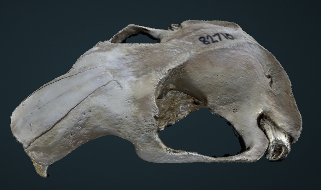
Super cool stuff. So what did we do? Well, I met Murat’s lab and his suite of postdocs and research tech: Dr. Chi, Dr. Sara, Dr. Rachel, and Sophie. In addition to speaking with Murat about upcoming potential collaborations, I worked most closely with Chi on mouse skull reconstruction, helping to refine techniques for that. You may recall some of my play with skull reconstructions in early 2020, both on larger and smaller mammals. Well these folks are doing it for real and for science, so it was fun to dive in.
This work with photogrammetric scans of samples is important for many of the reasons ODM is important in other contexts: scanning can be exceedingly expensive to do at scale with X-Ray approaches. Approaches like photogrammetry both augment those deeper approaches with surface textures and can also can be done with much less expensive set-ups. We have experience with this driving broader use and adoption as well as application to a broader set of samples (imagine scanning an entire set of samples from a natural history museum collection).

I won’t get into too much detail until our paper led by Chi and Murat is finished, but we refined his process with some small tweaks to data collection process and data processing settings to get some best in class results both to get best possible detailed meshes as well as repeatable measurable outputs.
In addition to working with Chi, It was also super interesting to talk to Rachel about her work sorting out the logistics of fetal mouse scans for developmental studies and get a snapshot of what Sara and Sophie are doing with respect to scan segmentation, deep learning, and better methods for delineating and understanding morphology now and in the future.
I’ll be posting more on these collaborations as they unfold. I can’t wait to continue.
